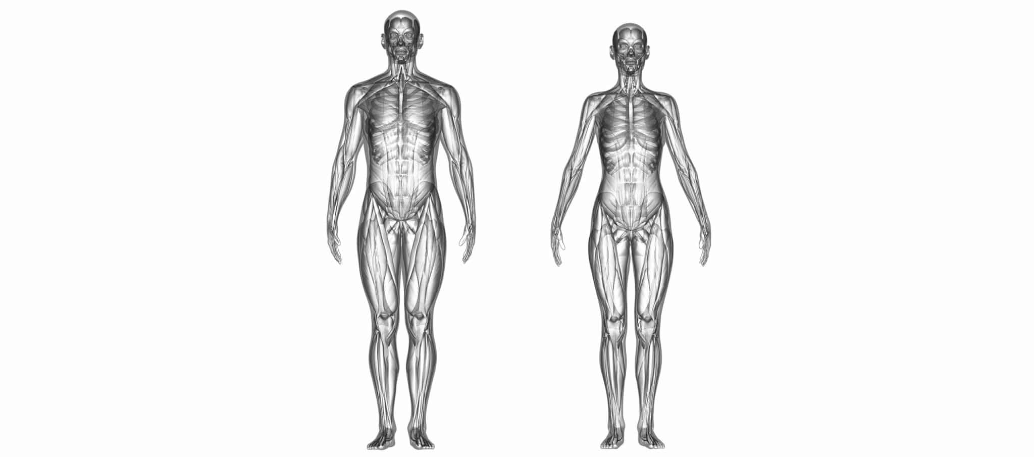
Tendon Anatomy
Tendons are cords composed primarily of collagen fiber; some are covered with a sheath that can produce a lubricant (synovial fluid) facilitating movement. One end is an extension of the muscle and the other is attached to the bone in the joint. They are resistant, but much less elastic than ligaments, and are part of the muscle structure. However, they do not contract like muscles. In addition, they are poorly vascularized so do not heal well when damaged, and are very innervated, which means they are extremely reactive to changes in pressure or direction exerted on a joint.
Their main role is to transfer the power generated by the muscles to the bone structure resulting in movement. However, they also play a very important intermittent role in joint stabilization. The ligaments are the primary, permanent stabilizers, but when they are pushed too far due to a brutal impact or a sharp or unusual movement, the tendons, due to their reactivity, can absorb these different mechanical traumas and thus prevent a fall or an injury (luxation, sprain…).
Tendon disorders
DIFFERENT TYPES OF LESIONS
Tendinosis is chronic tendon damage caused by repeated micro-traumas resulting in the formation of scar tissue (cystic nodules or calcinosis) due to micro-tears in the fibers. This mechanism induces tendon stiffness and fragility, which can sometimes result in partial or complete rupture. It is regularly observed in athletes, but can also occur in sedentary subjects, in particular the elderly.
Tendonitis is inflammatory damage caused by a rubbing mechanism, which generally induces excess secretion of synovial fluid, tissue inflammation and sometimes, with more advanced forms, adherence of fibers with other tissues. It is characterized by pain, edema, redness and a warm or even burning sensation.
Trauma injuries are caused by violent impacts or sudden, sharp changes in direction. They can lead to rupture or luxation. They are generally associated with other damage around the joint (bone fracture, sprain).
DIFFERENT CONDITIONS
Conditions implicating the ends of the tendons
Insertion disorders: myotendonitis or tenomyositis is the inflammation of the junction between the muscle and the tendon. The pain often develops progressively during effort. Enthesitis or tenoperiostitis is damage where the tendon inserts into the bone. The pain is generally very localized and stretching the tendon is painful.
Tendino-bursitis is the inflammation of the end of the tendon and the bursa located on the outside of the joint (between the bone and the tendon). The synovial fluid secreted by the bursa facilitates movement.
Tendinopathies
Real tendonitis: This is the inflammation of the tendon. It is painful on palpation, during passive stretching or forced movement.
Tenosynovitis: This is the inflammation of the synovial tendon sheath.
Tendinosis: This is a chronic degenerative tendon injury linked to repeated microtraumas as a result of overuse or age. It can cause premature wear of the tendon, which can lead to tendon rupture in severe cases.
Partial or complete tendon rupture: This is due to a trauma or worsening of tendinosis. The pain is often sharp and generally results in immediate functional incapacity. The subject may feel and even hear a snap when the injury occurs.
Tendon luxation: Often as a result of a trauma, it is when the tendon slips out of its normal anatomical position.
Causes and contributing factors
Tendon injuries are generally linked to tendon overuse, which is why athletes are often affected. Some sports such as football, skiing, basketball, or tennis that require abrupt changes in direction or stopping suddenly are particularly risky as they put the joints under intense strain.
However, morphological defects inducing tendon hyperstimulation (hallux valgus), repeated movements that are not necessarily very physical, or simply poor posture can exacerbate these injuries.
Some jobs are more particularly susceptible to this type of damage to the upper limbs (cashiers, violinists, prolonged work on a computer…).
The risk is greater in athletes due to insufficient warming up, changes in surface or equipment, training that is too intense and too long, climatic conditions (low temperatures), poor posture or when resuming training after a period of rest.
In the case of tendonitis, factors such as infection, diet or dehydration can lead to high concentrations of uric acid often implicated in this type of inflammatory damage.
Treatment
The aim is to relieve the pain and reduce the inflammation. Oral analgesics are often recommended. Oral or local anti-inflammatories can also be recommended. For inflammatory damage, it is recommended to put ice on the injury.
Therapy must include partial or total rest of the limb in question. Functional rehabilitation and physiotherapy are generally recommended.
The cause is also investigated to prevent any relapses: poor posture at work inducing over intense stimulation of the tendon, unsuitable footwear, uneven or slippery ground, genetic or acquired malformations…
Tendons are poorly vascularized and so do not heal well. Recovery using conventional treatment methods is rarely completely satisfactory, and relapses are common.
Surgery is only used if conventional treatment fails, with chronic, painful damage that impairs daily activities, in athletes, or when there is no alternative. It can consist in removing the scar tissue, suturing the tendon following a partial or complete rupture, or even attaching the tendon to the insertion site in the bone. Surgery enables lasting healing of tendons; however, as with any surgical procedure, there is a risk of inflammation, infection, and thromboembolism, as well as a risk linked to the anesthetic. It is therefore necessary to discuss the benefits with an orthopedic surgeon.
Stem cell injections and PRP therapy (platelet rich plasma) are cutting-edge techniques that enable rapid and effective healing, but are currently only used in athletes.

