Revision hip replacement
You have a loosening of the hip replacement
You are going to undergo revision hip replacement

You have a loosening of the hip replacement
You are going to undergo revision hip replacement
Revision hip replacement
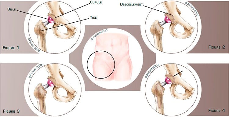
A total hip replacement is made up of two main components: the socket, which is a hollow hemispherical component implanted in the acetabulum, and the femoral stem with a ball, which is implanted in the femur. The ball replaces the head of the femur and articulates with the socket (figure 1).
With time, and in particular with implants fitted in the past, the friction surface between the two components wears. The debris from this wear is released around the implant in the form of micro-particles.
The body recognises these micro-particles and tries to digest them. An inflammatory reaction occurs to destroy them, but involuntarily attacks the interface between the bone and the implant.
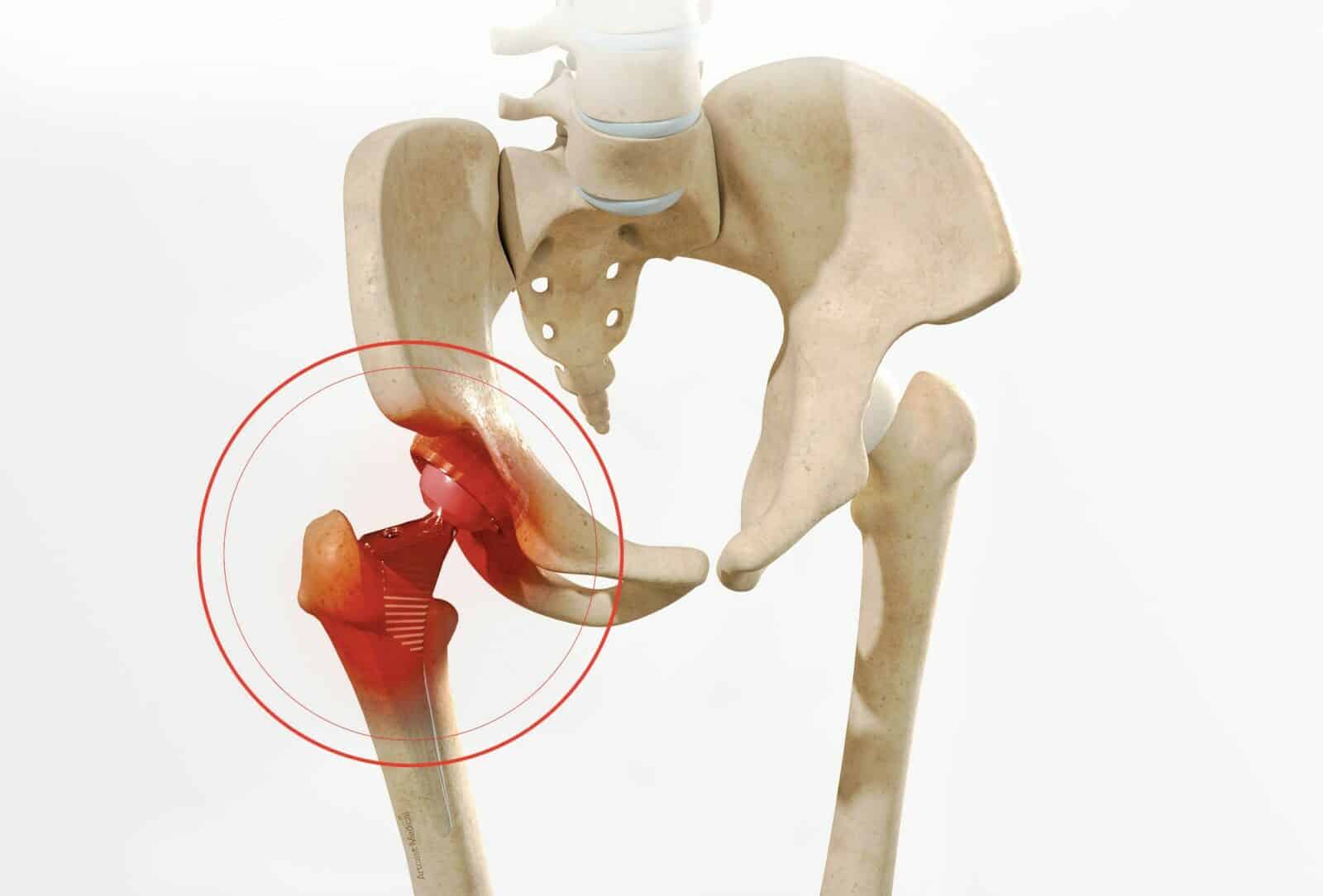
This leads to the progressive loosening of the implant resulting in mobility in relation to the bone. The loosening can concern the socket (figure 2), the femoral stem (figure 3), or both (figure 4). Causes other than natural wear, such as a chronic infection, poor positioning of the implant, or recurrent dislocations, can result in this developing more rapidly after the operation.
The loosening will cause pain, limping, even a shortening of the limb, as well as stiffness, progressively decreasing the functional possibilities of the implant.

The loosening as well as the inflammation around the implant will get increasingly worse, causing greater bone pain. The bone around the implant will get thinner and thinner and may fracture. All these phenomena are responsible for an increasingly painful discomfort. An x-ray, scan or bone scintigraphy will confirm the diagnosis. Once the diagnosis has been confirmed, the implant must be changed. The objective of the operation is to prevent the deterioration of the bone as early as possible and thus relieve the pain, recover mobility and return to normal walking.
The aim of the operation is to remove and replace the loosened component(s) of the old implant. In most cases, the replacement is said to be simple, that is, there are no other procedures associated. The faulty implant is extracted easily and the debris due to wear is cleaned. According to the seat of the loosening, a new socket (figure 5), a new, longer femoral stem (figure 6), or a new implant is fitted (figure 7).

If some of the areas of the bone around the prosthesis have been destroyed by the inflammation,
reconstruction by bone grafting will be necessary at the same time. The graft can be taken directly from the skeleton, or from a donor once treated in the laboratory.

On the acetabulum, after cleaning the debris and the faulty bone, the bone is rebuilt using a bone graft and supported by a metal structure. A socket can thus be fitted in the right conditions (Figure 8). An identical procedure is carried out on the femur. A femorotomy, or an opening down the side of the femur, is sometimes required to remove the stem. It also enables better bone regeneration. A long stem is fitted and stabilised using screws in the distal part. Metal rings are also positioned around the femur to hold it in place (figure 9). If the loosening concerns the socket and the femoral stem, these two procedures are carried out at the same time (figure 10).

If the loosening is linked to a chronic infection, a period of approximately 6 weeks is often required between the removal of the old implant and the placement of the new one. During this period, a Spacer, or temporary implant, is fitted to leave the bone time to heal with antibiotics. In the case of recurrent dislocations or in the elderly, a dual mobility implant can be recommended (figure 11). The ball is a large dual mobility head giving it additional stability. The operation lasts about 2 to 3 hours on average, and requires around 7 days in hospital. The operation can be carried out under spinal or general anaesthesia. Your anaesthesiologist will decide with you the best type of anaesthesia according to your state of health. After the operation, the incisions are covered with a sterile dressing, which is left in place for 10 days. The pain will be managed and monitored very closely during the post-operative period, and the treatment will be adjusted accordingly.
In the case of a simple replacement, the physiotherapist will get you up the day after the operation and help you to walk. Walking sticks may be useful to start with. You can go up and down stairs within 3 days. Except for very specific cases, it is not necessary to go to a rehabilitation centre. You physiotherapist will carry out your rehabilitation. Driving can be envisaged rapidly, and you can generally return to work after the 1st month, depending on your profession; office work can be sooner. A progressive return to gentle sports activities can generally be envisaged after the 3rd month.
When bone reconstruction is carried out at the same time, you will have crutches to help you move around for 6 weeks so as not to put too much weight on the hip in the case of the acetabulum and for 2 and a half months in the case of the femur. You may need to go to a rehabilitation centre after the operation. Driving can be envisaged after the 2nd month in the event of a reconstruction of the acetabulum and after the 3rd month for the femur. You can generally return to work after the 3rd month, depending on your profession; office work can be sooner. A progressive return to gentle sports activities can generally be envisaged after the 6th month.
In addition to the risks associated with any surgery and the anaesthetic, there are some risks specific to this surgery :
This list of risks is not exhaustive. Your surgeon can provide you with any additional explanations and will be available to discuss the advantages, disadvantages and risks of each specific case with you.
The results of this technique are very encouraging as an often spectacular disappearance of the pain along with a rapid recovery of mobility and muscle strength are observed. Even if the result is impressive and that many patients forget they have an implant, it is however preferable to avoid physical work and violent sports. These activities can increase the wear and decrease the lifespan of the implant. Some activities such as cycling, swimming, golf or walking are possible, and even recommended, whereas care should be taken with skiing, tennis and jogging. The average lifespan of a knee replacement is about 20 years. With the progress in the materials used today, we hope that the results and the longevity will continue to improve.
Laissez votre commentaire