 Doctor Philippe Paillard
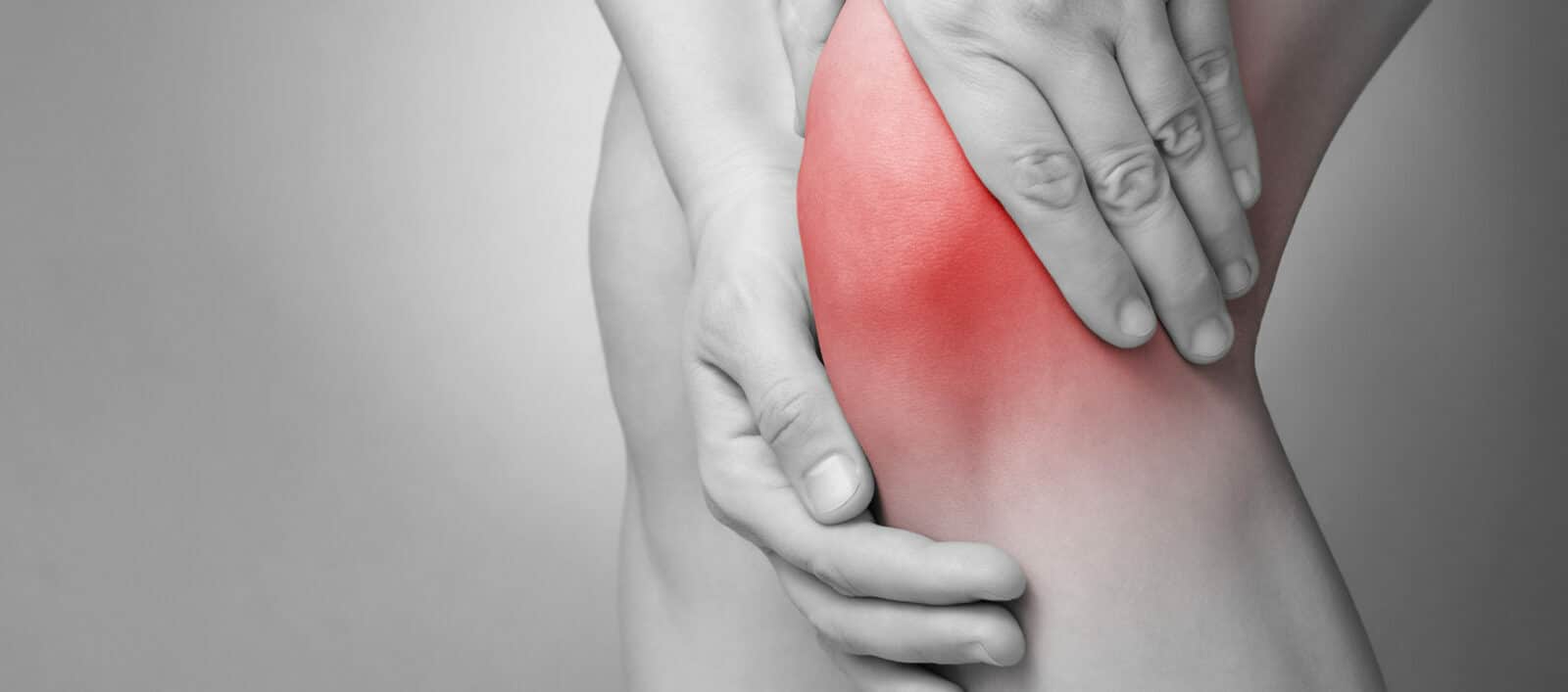
General information
The knee is a complex, weight-bearing joint located at the intersection between the leg and the thigh and is always under strain as it supports the weight of the body and enables movement. Its primary motion is flexion/extension. It comprises the ends of the femur and the tibia, and the patella, and is held together by a powerful stabilizing mechanism comprising ligaments and tendons.
The patella is located at the front of the knee and is connected to the quadriceps at the top and the tibia at the bottom by tendons. Two ligaments, the medial and lateral patellar retinacula, provide lateral stability.
The patella is a sesamoid bone, a bone that develops in a tendon and is found in some joints, but due to its size, it is generally considered as a bone in its own right. Its role is to protect the quadriceps tendon and increase the power transferred to the tibia during extension: when the quadriceps, the most powerful muscle in the body, contracts, it pulls the patella upwards, which in turn pulls the tibia thus resulting in leg extension.
As with other joint bone surfaces, the patella is covered with cartilage; this ensures smoothness of movement and protects the underlying bone tissue.
Mechanisms and causes
Patella pain occurs when there is an imbalance between the quadriceps and the hamstring (group of muscles and tendons, prime leg flexor and quadriceps antagonist, provides opposing force). This imbalance can occur due to:
- Hamstring stiffness: the quadriceps must therefore increase the power transferred to the patella to straighten the leg.
- Loss of quadriceps muscle resulting in abnormal pressure on the patella.
In both cases, abnormal rubbing of the joint surface can ultimately result in cartilage damage called chrondomalacia.
Various factors can increase this risk:
- In athletes: over intense training, resuming training after a period of rest or convalescence (resulting in quadriceps muscle wasting), absence or lack of stretching (causing hamstring stiffness), wearing shoes that are unsuitable for the activity in question or the terrain.
- In adolescents: morphological changes linked to growing, sudden increase in weight.
- In the entire population: changes in pace and activities (elevator out of order), occasional leisure activities (especially skiing), traumas (requiring convalescence resulting in quadriceps muscle wasting), carrying heavy loads (when moving house, for example).
- Women seem to be more exposed to this dysfunction.
Symptoms
Pain can be experienced during exertion and at rest, but is often hard to locate. It is generally felt at the front of the knee behind the patella, but can also be felt around the joint and may even radiate to the back. It is generally experienced during flexion: sitting for long periods, crouching, going down stairs, difficulty getting up without using the upper limbs.
The pain can be unilateral or bilateral, and can be felt long before irreversible cartilage damage appears. Inversely, some patients do not feel any pain even though the cartilage has already deteriorated.
Other symptoms can be associated with patella pain:
- Edema: abnormal rubbing can cause excess secretion of synovial fluid (joint lubricant) resulting in joint swelling.
- Instability: this is due to the slackening of the quadriceps as a result of the pain. In this case, there is no dislocation of the patella, but more a feeling of giving way.
- Blocking: feeling of stiffness or limited flexion due to the pain. This blocking can increase the loss of quadriceps muscle tone and therefore the imbalance.
- Grating, cracking, snapping can be heard without systematically being synonymous of severity.
Diagnosis
Diagnosis is above all based on the clinical examination and the interview, and often requires the skills of specialists.
Imaging such as an x-ray or an MRI, is sometimes necessary to eliminate other diagnoses such as ligament or meniscal damage.
An arthroscopy, performed under anesthetic in an operating room, can also be carried out to assess the condition of the cartilage. It is generally performed at the same time as the operation to restore the cartilage, when this is possible.
The diagnostic phase is very important to analyze the muscle mass as well as the origin of the imbalance to help determine the treatment.
Treatment and convalescence
As with any episode of pain, the initial treatment is rest. It is necessary to restrict activities, avoid painful movements, and possibly wear a knee brace to relieve the joint.
Anti-inflammatories, analgesics and cryotherapy (application of cold) can help relieve the pain.
Treatment is then mainly physiotherapy -based and consists in increasing quadriceps muscle tone and loosening the hamstring with regular stretching.
However, the treatment must also look into the causes and factors to help reduce relapses. Arthroscopic surgery can be performed to repair advanced patellar cartilage damage. However, as it is rather hard to treat, the damage should be treated before it becomes irreversible.