 Doctor Philippe Paillard
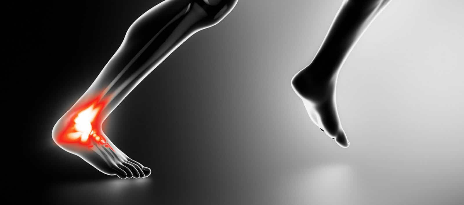
GENERAL INFORMATION
The heel is the anatomical structure aligned vertically with the leg at the back of the foot. It comprises two bones, the astragalus (or talus) and the calcaneum (the biggest bone in the foot), as well as muscles, ligaments, tendons, nerves… which enable it to function. Due to its location, the heel sustains most impacts (initial contact when walking, landing…) and is therefore under a lot of strain, even when the person does not do any particularly physical activities. Trauma, overuse, or certain conditions can damage different parts of the heel resulting in pain.
CAUSES
Intense sport, repeated movements, regularly carrying heavy loads or being overweight (excess weight, obesity or pregnancy), congenital or acquired deformities, wearing shoes that are worn or ill-adapted to the subject’s morphology or the activity in question, and aging can all lead to mechanical dysfunction or traumatic injuries causing heel pain.
Tendonitis
Inferior heel pain could be caused by tendonitis of the plantar fascia tendons. Posterior pain is usually Achilles tendonitis. In both cases, it can be insertional tendonitis (inflammation of the tendon is thus around the calcaneum) or tendino-bursitis (inflammation of the end of the tendon and the bursae located between the heel bone and the tendon).
Tendon damage
Inferior heel pain can be linked to plantar fasciitis, or the inflammation of the plantar fascian ligaments connecting the toe bones to the calcaneum. Avulsion fractures or the formation of a calcanean bone spur, also called “Lenoir Spur” – a bone excrescence – can occur during violent traumas or with advanced forms.
The calcaneum articulates with the astragalus, located above. As in any joint, the bones are covered with cartilage. Due to natural aging, overuse or a trauma, cartilage damage is likely to develop into osteoarthritis. When it concerns this part of the anatomy, it can cause heel pain.
Calcaneum fractures
It can be so-called stress fractures, but are more generally related to violent traumas and particularly landing on the heels following a fall. When this happens, the calcaneum is compressed between the astragalus (which bears the weight of the body, increased here by the fall) in one direction and the ground in the other. This generally results in comminuted fractures, which are complicated and longer to treat, notably when the various fragments move.
Deformities
Some congenital or acquired deformities can also be the direct cause of this type of pain. The most common are architectural disorders of the feet (flat or arched feet), calcaneum spurs (seen previously), but also morphological anomalies not directly related to the foot such as knee deformities (valgum or varum), or unequal leg length.
Aging
It can lead to vulnerability and stiffness of the tendons and ligaments, fragility of the bone structure, but also thinning of the fat layer located under the calcaneum. This “cushion” helps absorb impacts, but it diminishes with age and can result in heel pain, often symmetric.
Mechanical dysfunctions
Various conditions can also be the direct cause of heel pain, or generate mechanical dysfunctions such as seen previously.
- Ankylosing spondylitis : Associated with other symptoms, heel pain is characteristic of this inflammatory disease and is sometimes one of the first signs indicating this diagnosis.
- Rheumatoid arthritis is an inflammatory joint disease. It can be located in different joints in the skeleton. When it affects the talocalcaneal joint, it can cause heel pain.
- Gout, usually causing pain in the big toe, is a chronic metabolic disease characterized by the deposition of uric acid crystals in different parts of the body. When the joints are affected, we talk of gouty arthritis. This disease is likely to cause heel pain when it affects the talocalcaneal joint.
- Server’s disease is osteochondritis of the foot (growth disorder involving bone and cartilage) affecting adolescents between the age of 10 and 16. Not all the causes are known precisely, but they can be directly linked to intense sports activities. It can cause micro-traumas of the calcaneum and inflammation of the Achilles tendon. It comes and goes with one-off or repeated flare-ups, characterized by heel pain, limping and sometimes joint blocking. The disease regresses and disappears when the child stops growing.
- Peripheral nerve disorders such as nerve compression, sciatica, peripheral neuropathies, whatever the cause (diabetes, Guillain-Barré syndrome…), can also result in heel pain.
- Psoriatic rheumatism, which affects the joints, can sometimes develop in subjects with psoriasis, a chronic inflammatory skin condition. The disease primarily affects the feet, especially the toes and the heels.
- Arterial disease, characterized by a loss of blood flow to the affected areas, it can also cause heel pain when it affects the lower limbs, resulting in burning sensations.
DIAGNOSIS
Heel pain is not always a specific indication of a disorder or trauma, so it is possible to rest the foot, apply ice and take analgesics or anti-inflammatories. However, if the pain persists, returns or seems sharper, causes aggravating posture or mechanisms (limping, standing on tiptoes), or there are other symptoms: higher body temperature, signs of inflammation (redness, heat, edema), skin damage, pain in other parts of the body, etc., it is recommended, child or adult, to consult a physician immediately.
The diagnosis is above all clinical and may require the skills of a specialist. The practitioner will assess the type of pain, when it occurs, triggering factors and location. However, in the absence of any related trauma or condition, it may be complicated to establish a diagnosis and further examinations may be required:
- X-ray : screening for fracture or deformity
- MRI : examination of the different parts of the heel
- Bone scintigraphy : bone analysis
- Ultrasound : visualization of the soft tissue (plantar aponeurosis, tendons, muscles…)
- Electromyogram : examination of a possible neurological affection
- Doppler : evaluation of
A blood test will provide precious information, in particular to clarify the diagnosis of an inflammatory or infectious disease.
All these tests will enable the right treatment to be initiated, as well as the causes to be defined in order to prevent any relapses.
In addition, heel pain may be the first sign of an underlying inflammatory disease (e.g. ankylosing spondylitis) or indicate a complication with regard to a pre-existing chronic disease (e.g. diabetes). A complete diagnosis will highlight these more serious conditions and enable the most suitable treatment to be initiated.
TREATMENT
Treatment of heel pain varies as there are numerous causes. The primary objective is to relieve the pain, but may also involve treating the causes in order to prevent relapses (typical in the case of sciatica, for example) or any possible subsequent complications.
Significant mechanical pain, when it does not result in any complications, is treated simply with painkillers or anti-inflammatories, rest and possibly cryotherapy (applying cold to the painful area). This is the case when it is related to tendonitis or Server’s disease, for example.
Other problems such as fractures or tendon or ligament ruptures may require surgery, which is generally followed by a period of convalescence of varying duration.
Others may be part of a broader treatment plan, which will never neglect the pain, but will look more particularly at treating or stabilizing a chronic or inflammatory condition.
Physiotherapy initiated immediately or following surgery is also an effective way of relieving the pain. However, the treatment of more general factors that can be directly implicated such as excess weight or poor footwear must not be overlooked.