Causes
Collarbone fractures often occur following a trauma and can occur at any age.
In children, adults and the elderly, they generally occur during a fall. In babies, it is generally a complication when maneuvering the shoulders during childbirth.
Collarbone fractures most commonly occur during indirect traumas as a result of landing badly on a hand, arm or shoulder. The force of the impact is transmitted to the clavicle, which then fractures. This is a common injury sustained by cyclists or horse-riders
Fractures caused by a direct impact are rare. They can occur during accidents due to a direct blow to the clavicle or wearing a seat belt. This type of injury is more likely to occur during rugby and fighting sports.
Pathologic fractures linked to tumors are specific cases that will not be addressed in this article.
Different types of fracture
Different lesions
- Simple fractures: one single fracture line with anatomical alignment maintained.
- Fracture with displacement: one or several fracture lines with rupture of the anatomical alignment
Different locations
- Diaphyseal (midshaft) fracture: this is the most common lesion and represents approximately 75% of collarbone fractures.
- Fracture of the distal third (shoulder side): this is the other type of fracture commonly encountered.
- Fracture of the proximal third (thorax side): this is very rare.
Diagnosis
Symptoms
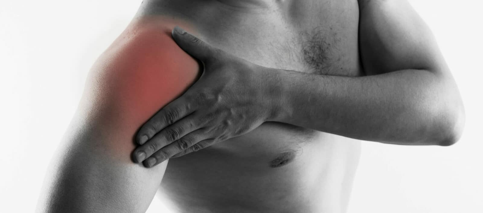
As with all fractures, the main symptoms include pain when touched or moved, as well as the rapid appearance of edema (swelling) or even bruising around the lesion.
It also results in partial functional impairment thus restricting normal movement.
These fractures can sometimes cause the shoulder to sag forwards or induce a change in the normal anatomical structure.
A “bump” can be seen in the middle of the bone in the case of diaphyseal fractures with displacement.
Additional examinations
-
- An x-ray is performed to confirm the clinical diagnosis and to accurately locate the fracture, as well as to determine the severity (extent of the displacement, for example).
-
- A scan is not always necessary. It is used to reinforce the diagnosis to help determine the best treatment..
Immediate complications
Collarbone fractures are generally minor fractures that do not result in any major complications. When faced with the aforementioned symptoms, the arm should be bent at 90° and held against the chest in a “sling”. The patient must then go to ER. However, some more serious cases may require more rapid treatment.
- Open fractures : These can occur in the event of diaphyseal fractures with displacement, when the internal fragment breaks through the skin. This type of fracture is not life threatening, but it does require rapid surgical treatment, especially with regard to the risk of bleeding.
- Neurological damage : The brachial plexus (group of nerves located in the shoulder region) can suffer damage during violent traumas, resulting in hyposensitivity and/or functional impairment of the upper limb.
- Vascular damage : Collarbone fractures, particularly in association with fractures of the first rib, can result in damage to the subclavian arteries – which is generally identified by the absence of pulse at the wrist – or the subclavian veins. Surgery is required rapidly to limit the risk of complications linked to bleeding.
- Pulmonary damage : With fractures resulting in significant displacements, the bone can pierce the lung causing a “pneumothorax”. This can lead to serious respiratory complications and be life threatening.
Treatment
Treatment can be surgical or not according to the type of fracture and the severity of the lesion. The outcome of collarbone fractures is generally favorable and sequelae are rare.
Non-surgical treatment
As it is non-invasive, this treatment is quite advantageous. However, it is only possible with simple diaphyseal fractures.
It consists in wearing a brace or “figure-of-8 bandage” to hold the shoulder back and maintain the anatomical alignment for the 6 to 10 weeks necessary for the bone to form a “callus” (heal).
After 3 months, if no healing is observed on the x-ray, then surgery will be necessary.
Surgical treatment
Surgery is indicated for diaphyseal fractures with displacement, and proximal or distal fractures.
It consists in a “reduction” of the fracture – recreation of the initial anatomical alignment of the bone – then an “osteosynthesis” – positioning of a plate and screws, or even artificial ligaments to hold the bone fragments solidly in place during healing. A brace must also be worn to hold the arm against the body for the first six weeks after the operation.
A second operation (approximately one year later) will be performed to remove the hardware. The bone will generally be temporarily weaker due to the holes left by the screws, but will heal within a few weeks.
Physiotherapy
Rehabilitation after healing is recommended whatever the treatment, as immobilization of a limb systematically leads to muscle atrophy. Different physiotherapy exercises will help restore circulation and strengthen the shoulder.

